WHAT IS OSTEOARTHRITIS?
Osteoarthritis (OA) is the most common type of arthritis.
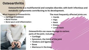
The earliest research on osteoarthritis was that it is degenerative diseases. current research has shown that it is a multifactorial and complex disorder, with both infectious and metabolic components contributing to its development.
Experts estimate that osteoarthritis affects more than 80% of persons over the age of 55, even if some never develop symptoms.
Around 60% of patients with osteoarthritis have symptoms that they can observe or feel.
In healthy person
The ends of the bones in your joints are covered with a smooth, slippery tissue called cartilage.
This cartilage cushions the bones and allows them to glide effortlessly when the joint moves.
Acting as both a shock absorber and a lubricant, cartilage ensures that the bones in your joints move smoothly and safely past each other.
In Osteoarthritis
The protective cartilage that cushions the ends of bones wears out and becomes rough. It causes discomfort, stiffness, edema, and restricted movement, particularly in the hands, knees, hips, and spine.
The main outcomes of OA are cartilage degradation, acute and chronic synovial inflammation, subchondral bone modification, the presence of osteophytes, and alterations in synovial fluid.
Osteoarthritis only affects joints, typically in the hands, knees, hips, neck, and lower back. It is the most common form of arthritis. There is no cure for osteoarthritis. It usually worsens slowly. While there is no cure, controlling weight, staying active, and sticking to a treatment plan can help slow its growth and improve symptoms.
However, the damage to the joints cannot be reversed. Staying active, maintaining a healthy weight, and receiving some therapies may decrease disease development and improve pain and joint function.
Related article: Rheumatoid Arthritis (RA): Symptoms, Causes & Risk Factors
CAUSES AND RISK FACTORS OF OSTEOARTHRITIS
- Age
- Obesity
- Joint injury
- Genetics
- Gender
- Sedentary lifestyle
- Joint overuse
- Musculoskeletal abnormalities
COMMON SYMPTOMS OF OSTEOARTHRITIS
- Pain
- Stiffness
- Swelling
- Limited movement
- Sounds
- Grating sensation
- Bone spurs
Related article: Everything You Need to Know About Osteoarthritis: Symptoms, Causes & Risks
HOW OSTEOARTHRITIS IS DIAGNOSED
There is no definitive test for osteoarthritis. To determine if you have osteoarthritis, consult your provider:
- Will observe the symptoms and medical history
- Perform physical examination
- Use X-rays or other imaging tests to look at your joints
- Lab tests to make sure that a different problem isn’t causing your symptoms
During a physical examination, your healthcare provider will assess the affected joint for tenderness, swelling, and range of motion.
Imaging tests may also be used to evaluate the joint in more detail.
X-rays are commonly performed to visualize joint structure, while MRI (magnetic resonance imaging) or CT scan (computed tomography) scans may be used to provide more detailed images of the bones and soft tissues.
Imaging tests
To get pictures of the affected joint, your healthcare professional might recommend:
X-rays test
X-rays are used to show the extent of joint deterioration, such as narrowing of joint space, which indicates cartilage loss, thinning or erosion of bone, often seen in more advanced arthritis, bone spurs (osteophytes) or other abnormalities, which develop as the joint tries to repair itself.
They also help doctors differentiate between different types of arthritis, such as osteoarthritis and rheumatoid arthritis, based on the pattern of joint changes.
Magnetic resonance imaging (MRI)
An MRI uses a strong magnetic field and radio waves to create detailed images of bones and soft tissues, including cartilage. While MRI is not usually required to diagnose osteoarthritis, it can be helpful in complex cases to provide additional information about joint structure and damage.
Lab tests
Analyzing blood or joint fluid can assist confirm the diagnosis.
Blood test- Although there is no blood test to diagnose osteoarthritis, certain tests can help rule out other causes of joint pain, such as rheumatoid arthritis.
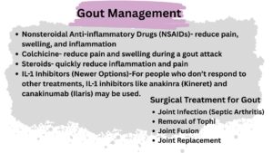
Analysis of joint fluids- A needle may be used to extract fluid from an afflicted joint. The fluid is then examined to see if your pain is caused by inflammatory arthritis, such as rheumatoid arthritis or gout, or an infection, rather than osteoarthritis.
TREATMENT AND MANAGEMENT OPTIONS
NON- PHARMACOLOGICAL MANAGEMENT
Early, nonsurgical treatment can help maintain joint mobility, improve strength, and relieve pain. Most treatment programs combine lifestyle modifications, medication, and physical therapy.
Lifestyle Changes includes
Exercise
Exercise can help improve physical function, reduce pain, and relieve stiffness. Moving your joints regularly strengthens the muscles around them, which supports joint health.
Low-impact activities such as swimming, water aerobics, cycling, and weight training are especially beneficial. Your healthcare provider may recommend consulting a physical therapist for guidance.
It’s also helpful to shift from high-impact activities—like running, jumping, or competitive sports—to low-impact exercises such as stretching, walking, swimming, or cycling.
Maintain a Healthy Weight
Maintaining a healthy weight can help reduce pain, particularly in weight-bearing joints such as the spine, hips, knees, and ankles. If needed, losing weight can lessen the stress on these joints and improve overall joint function.
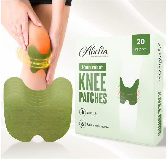
Heat and Cold Therapy
Applying heat or cold to affected joints can help relieve pain and stiffness. Your healthcare provider can guide you on how often and for how long to use a heating pad, ice packs, or a cool compress.
Heat therapy not only reduces discomfort but also increases the expression of heat shock protein 70 (HSP70), which has a calming effect. HSP70 plays a role in protecting cartilage, reducing inflammation, and preventing chondrocyte apoptosis.
Cold therapy (shallow cold applications) primarily provides pain relief, though it does not offer the same cartilage-protective benefits as heat therapy.
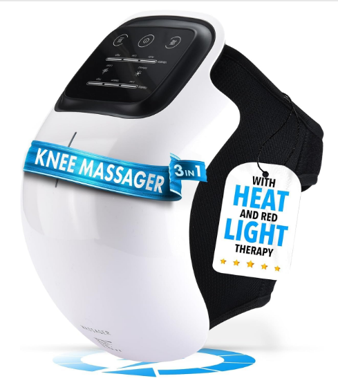
Pulsed electromagnetic field therapy (PEMF)
PEMF is a non-invasive treatment that uses low-frequency electromagnetic waves to stimulate and heal the body’s cells, tissues, and bones.
PEMF therapy may:
- Reduce joint pain and stiffness
- Improve mobility
- Slow cartilage degeneration
- Support natural tissue repair
Note: Not recommended for people with pacemakers, pregnancy, or active bleeding disorders. Always consult a healthcare professional before starting therapy.
Low-level laser therapy (LLLT)
LLLT, also known as cold laser therapy, is a non-invasive treatment that uses low-intensity laser light to stimulate healing in tissues without producing heat or damaging the skin.
LLLT combined with exercise has been shown to alleviate pain while improving mobility and movement in patients with knee OA.
LLLT can:
- Decrease joint pain and swelling
- Improve mobility and flexibility
- Promote cartilage repair and reduce joint inflammation
It is often used alongside physical therapy, medication, or exercise for better results.
Radiation also increases local microcirculation and is highly recommended as a supplement to other treatments for OA.
Massage
Massage for 60 minutes per week after eight weeks of therapy improved pain management. Massage improves blood flow and relaxes muscles around the affected joints. It helps reduce pain signals and enhances flexibility, especially in knees, hips, and hands.
Better blood circulation delivers oxygen and nutrients to joints. It helps clear out inflammatory waste products, reducing swelling. By decreasing pain and relaxing the body, massage often leads to better sleep and emotional balance.
Note: Always consult a physiotherapist or certified massage therapist for proper technique.
Supportive devices
Wearing shoe inserts or a brace can support and stabilize your joints. Using a cane or walker can take pressure off your affected joints and help you move safely.
Complementary therapy
Complementary therapies may work alongside other treatment options. Examples: acupuncture, massage, meditation, tai chi and dietary supplements.
Talk to your provider before you start taking any herbal or dietary supplements.
PHARMACOLOGICAL MANAGEMENT
Nonsteroidal anti-inflammatory drugs (NSAIDs) and analgesics
NSAIDs, such as ibuprofen and naproxen, are commonly used to reduce inflammation and relieve joint pain.
Other oral analgesics, including acetaminophen, diclofenac, and COX-2 inhibitors, may also be prescribed.
In some cases, intra-articular corticosteroid injections are used to alleviate arthritis-related discomfort.
Topical Agents
Topical treatments for arthritis include lidocaine, NSAIDs, and capsaicin. Applying NSAIDs topically allows the medication to be absorbed locally at the affected joint, reducing pain while minimizing systemic side effects compared to oral medications.
Intra-articular therapy
Sometimes, when a joint is very painful or swollen, a doctor can inject a strong anti-inflammatory medicine called a corticosteroid directly into the joint. This helps reduce pain and swelling for a short time.
It doesn’t cure the arthritis but helps you feel better temporarily. OA is also treated with corticosteroid injections into articular cartilage.
Example: Methyl prednisolone acetate, betamethasone acetate/betamethasone sodium phosphate, triamcinolone acetonide, triamcinolone hexacetonide, and betamethasone dipropionate/betamethasone sodium phosphate.
Intra-articular therapy helps in
- Reducing inflammation
- Relieving pain and swelling
- Improving joint mobility
Anti-cytokine therapy
Anti-cytokine therapy is an advanced treatment approach that targets specific inflammatory molecules (cytokines) responsible for joint damage and pain in osteoarthritis. It is part of the newer generation of biologic therapies designed to control inflammation at the molecular level.
Anti-cytokine drugs block the activity of these harmful cytokines, reducing inflammation and protecting cartilage from further damage. They work by neutralizing the cytokine or blocking its receptor so it cannot signal the inflammatory response.
Infliximab, golimumab, etanercept, certolizumab pegol, and adalimumab are five anti-TNF agents that have been documented for the treatment of rheumatoid arthritis. Tocilizumab is an anti-inflammatory medication that prevents the activation of the IL-6 receptor. Anakinra blocks the biological function of naturally occurring IL-1by competitively inhibiting IL-1 binding to IL-1R, which is expressed in a variety of tissues and organs.
Benefit:
- May reduce joint inflammation and pain
- Could slow cartilage breakdown
- Useful for patients not responding to conventional therapies
Limitations and Precautions
- Still under clinical research for osteoarthritis — not yet a routine treatment.
- Can cause immune suppression, increasing infection risk.
- Must be prescribed and monitored by a rheumatologist or specialist.
Omega-3 fatty acids as dietary supplements
Eicosanoids are substances in the body that control and regulate inflammation — they can either increase or reduce it. These eicosanoids are made from PUFAs (polyunsaturated fatty acids), which are essential fats our body needs but cannot make on its own.
When people take omega-3 fatty acids (a type of PUFA, found in fish oil and some plants), studies have shown that they reduce substances that cause inflammation and increase those that fight inflammation — both in laboratory and human studies.
In numerous animal models and clinical experiments, omega-3 fatty acids have been demonstrated to have anti-inflammatory properties.
Some studies have indicated that taking an omega-3 fatty acid supplement can considerably relieve OA symptoms in the knee cartilage and the lower region of the femur (a vertebra).
Herbs and Ayurvedic formulations
Several herbs have been utilized in Ayurveda for many years, and many of them are regularly used in human medicines; their usage in humans is thought to be entirely safe.
The condition is treated with the traditional Ayurvedic medications Triphala churna and Triphaghula, as well as Balaraja and Dashmoolasa.
Triphala churna: Triphala and its components contain anti-inflammatory, antioxidant, cytoprotective, and rejuvenating properties (Rashayana). It lowers inflammatory mediator levels and prevents lipid peroxidation.
Dashamoola: Dashamoola formulations contain the roots of ten plants that are also beneficial for vata-roga, as the name implies. Since ancient times, Dashamoolarishta has been utilized for its anti-inflammatory properties, as well as to relieve the inflammation and suffering associated with arthritis.
Note: One laboratory experiment of these analgesic combinations was demonstrated to reduce OA pain, while others were shown to decrease cartilage degeneration, a condition in which excessive Vata energy produces pain, immobility, and rigidity of the limbs.
SURGICAL MANAGEMENT
Most people do not require surgery to address osteoarthritis. If you have significant symptoms and alternative therapies have not been effective, your provider may propose surgery.
Surgery may be used if other therapies have failed to improve the patient’s symptoms. Arthroscopy, cartilage repair, knee arthroplasty, and osteotomy are surgical therapies for knee osteoarthritis.
The location, stage of OA, comorbidities, and people suffering on the opposite side all influence which of these procedures is best suited.
Arthroscopy
Arthroscopy is a minimally invasive surgical procedure that allows doctors to see inside a joint using a small camera
The surgeon makes tiny incisions near the affected joint (such as the knee, shoulder, hip, or wrist). A thin tube with a camera (arthroscope) is inserted through one incision. The camera displays detailed images of the joint on a screen. Through other small incisions, surgical tools can be inserted to remove damaged tissue, smooth rough cartilage, cysts, damaged lining, or repair ligaments.
Limitation
May not stop arthritis progression — it relieves symptoms but doesn’t restore lost cartilage.
Not suitable for severe osteoarthritis, where joint replacement may be needed.
Possible minor risks: infection, stiffness, or swelling.
Knee arthroplasty
Knee arthroplasty, commonly called knee replacement surgery, is a surgical procedure where a damaged knee joint — often due to osteoarthritis — is replaced with artificial components (prosthesis) to relieve pain and restore mobility.
Unicompartmental (partial) knee arthroplasty or unloading osteotomy. When osteoarthritis (OA) affects only one part (compartment) of the knee — usually the medial, lateral, or patellofemoral compartment — surgeons may consider less invasive alternatives to total knee replacement:
Only the damaged compartment of the knee is replaced with a prosthesis. The healthy parts of the knee are preserved. Less invasive than total knee replacement, with quicker recovery and better range of motion.
Total knee arthroplasty
In older patients with advanced knee OA, total knee arthroplasty is a routine and safe procedure. The surgeon removes damaged cartilage and bone from the femur, tibia, and sometimes the patella. These are replaced with metal and plastic prosthetic components designed to mimic the knee’s natural movement. The prosthesis restores alignment, stability, and function of the knee joint.
Osteotomy
Osteotomy is a surgical procedure in which a surgeon cuts and reshapes a bone to shift weight away from a damaged joint area, reducing stress and pain. It is often used in knee osteoarthritis when only one compartment of the joint is affected.
Limitation
- Recovery involves rehabilitation and physiotherapy
- Not ideal if multiple compartments of the knee are affected
- Surgical risks include infection, bleeding, or delayed bone healing
Joint fusion
Joint fusion, also called arthrodesis, is a surgical procedure in which the bones of a joint are permanently joined together to eliminate movement. This is done to relieve severe pain in a joint that cannot be managed by other treatments.
A surgeon eliminates the joint by fastening together the ends of bone (fusion). Pins, plates, screws, or rods may hold bones in place while they heal. This procedure eliminates the joint’s flexibility. Fusion is most commonly performed in the spine, hand, and foot.
Limitation:
- The fused joint cannot move, so mobility is reduced
- Stress may increase on adjacent joints, possibly causing arthritis there
- Usually considered a last-resort option when other treatments fail
Joint Replacement
In a joint replacement procedure (also called total joint arthroplasty), a surgeon removes damaged portions of the bone and replaces them with artificial components made of metal, plastic, or a combination of both. This helps restore joint function and reduce pain.
Total joint replacement is the most effective surgical treatment, with favorable patient outcomes for hip, knee, and shoulder replacements. Although there are numerous prosthetic devices available, no controlled trials have been conducted to compare them.
Most modern joint prostheses are predicted to last 15 to 20 years for the majority of patients. Other surgical therapies for osteoarthritis are available, but none have proven to be as effective as total joint replacement.
Available gaps and future therapies
In OA, cartilage in the joints breaks down over time, leading to pain, stiffness, and reduced mobility.
Certain enzymes, called matrix-degrading enzymes, contribute to this breakdown:
- MMP-13 (Matrix Metalloproteinase-13): Breaks down collagen, a key structural protein in cartilage.
- ADAMTS-5: Breaks down aggrecan, another essential component of cartilage.
Scientists studied OA in mice to test treatments.
They used CL82198, a drug that inhibits MMP-13 activity. They found that Blocking MMP-13 with CL82198 in mice cause:
- Protected cartilage cells
- Slowed cartilage breakdown
However, such findings in humans are yet to be verified.
Further research is needed to fully evaluate the efficacy and safety of MMP inhibitors.
Read article:
Osteoarthritis Symptoms, Cause and Risk Factors
Ginseng: Absorption, Benefits, and side effects
Everything You Need to Know About Osteoarthritis: Symptoms, Causes & Risks
Recommended Products